
Real-Time Neutron Diffractometer (RTD)
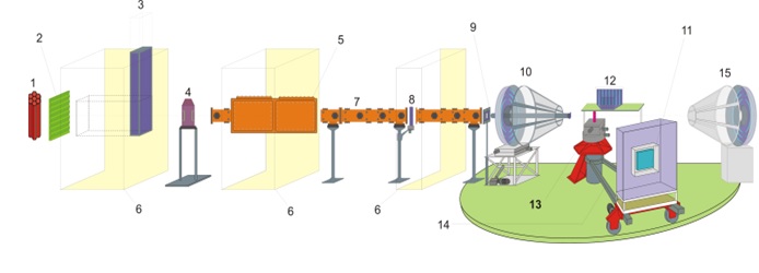
1 – reactor core
2 – moderator
3 – schieber (primary beam shutter)
4 – chopper
5 – beam splitter
6 – biological shielding
7 – mirror neutron guide
8 – secondary beam shutter
9 – adjustable diaphragm
10 – backscattering 8-ring detector
11 – position-sensitive detector
12 – 3 banks 8 3He “point” detectors
13 – goniometer and sample position
14 – PSD movable arm
15 – small-angle ring detector (in final stage of installation)
Instrument Responsible:
tel. +7 (49621) 6-62-82
Beskrovnyi Anatolii
tel. +7 (49621) 6-53-03
e-mail: This email address is being protected from spambots. You need JavaScript enabled to view it.
Research areas:
RTD spectrometer is a TOF diffractometer of medium resolution employed to study:
1. Crystal structures;
2. Magnetic structures;
3. Nanomaterials;
4. Phase transitions (including in real-time mode), crystallization and hydration-dehydration kinetics;
5. Chemical reactions (including in real-time mode);
6. Study of domain structure and diffuse scattering in single crystals;
7. Low-periodicity/long-period structures like lipid membranes;
8. Incommensurate and modulated structures. Possibility of registration of low-intensity superstructure peaks (~ 0.1-0.01% of the intensity of main reflections).
Basic parameters
| Neutron guide | Ni, mirror |
| Guide aperture | 15 mm × 180 mm |
| Moderator - sample distance | 23.85 m |
| Detector - sample distance | 0.15 - 2.0 m, changeable |
| Neutron flux at sample position | 5×10⁶ m/cm2/s |
| Range of wavelengths scattering angles d-spacing |
1.0 - 18 Å 1 - 170° 0.6 - 300 Å |
| Resolution, Δd/d, θ = 80°, d = 2 Å | 1 % |
| θ = 10°, d = 60 Å | 10 % |
Detector systems
The diffractometer is equipped with various types of detectors:
• 3 banks 8 detectors SNM-17 filled with 3He positioned at various scattering angles. They are usually used to study crystalline and magnetic structures of powders;
• Two-coordinate position-sensitive detector. Sensitive area – 225×225 mm. Resolution (Dx, Dy) ~ 0.2 cm;
• Ring detector with 8 concentric rings installed at large (“backscattering”) angles.
• Similar ring detector (but with azimuthal resolution) is in the process of installation at small angles.
Typical spectra
Diffraction spectra obtained at time-of-flight instruments can be conveniently represented as X-Y graphs, where the ordinate is interplanar distances and the abscissa is the scattered neutron intensity. To calibrate spectrometers, scattering from a standard sample is used. The energy (wavelength) distribution of the incident neutron beam is not uniform. Although it resembles a Maxwell distribution, its shape is modified by the shape of the neutron source pulse, type of moderator, form of a neutron guide, etc. To determine the shape of an incident neutron spectrum, the scattering from a vanadium sample is used.
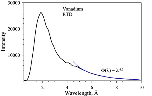
Vanadium spectrum representing the spectrum of the neutron beam at sample position. For λ > 5Å the intensity decreases as λ-3.2.
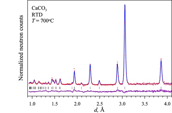
Normalized experimental spectrum of CaCO3 (red dots) at 700°C formed by thermal decomposition of C2CaO4 (H2O) (Calcium Oxalate Monohydrate) and the result of Rietveld refinement fit (solid blue line). Vertical bars are the calculated positions of diffraction peaks. The difference curve (experiment minus calculation) normalized to the root-mean-square deviation at a point is shown below (purple).
Sample environment equipment
The equipment used at the DN-2 spectrometer includes:
• Cryostat on the basis of closed-cycle helium refrigerator. Temperature range – 10 – 350 К.
• Water thermostat for investigations of biologic samples. Temperature range – from room temperature to 100 °С. Control of humidity.
• Vanadium furnace. Temperature range – from room temperature to 850 К.
• Muffle furnace for backscattering measurements up to 1400 K.
• Muffle furnace for small-angle measurements up to 1400 K.